Ileoanal Anastomosis Illustrations

Figure 1. Normal Gastrointestinal Anatomy and Physiology

1. Food enters the digestive system through the mouth.

2. Food travels the length of the esophagus.

3. Enters the stomach where the food mixes with digestive fluids.

4. Enters small intestine where digestive fluids continue to be secreted. The majority of digestion and some nutrient absorption takes place.

5. Enters large intestine or colon where water is absorbed and stool is stored.

6. Enters the rectum.

7. When patient feels the urge to pass stool, final passage of stool is through the anus.

Return to "What is a J-Pouch?What is J-Pouch Surgery?"


Figure 2, Stage 1: Ileoanal Reservoir Surgery

1.The large intestine (colon)is removed.

2. The lining of the rectum is removed but the sphincter mechanism is saved.

Return to "What is J-Pouch Surgery?"


Figure 3, Stage 1 continued: Ileoanal Reservoir Surgery


A temporary loop ileostomy is created to "detour" the contents of the small intestine so the newly created reservoir may heal.

Return to "What is J-Pouch Surgery?"


Figure 4, Stage 2: Gastrointestinal tract after Ileoanal Reservoir Surgery
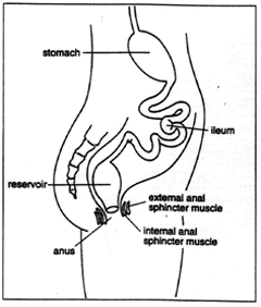


1. Approximately 2 to 3 months after the first surgery, the ileostomy is closed.

2. Food now enters the mouth, then into the stomach, then through the small intestine into the reservoir.

3. Waste is stored in the reservoir until the need to have a "bowel movement."

Stool is then passed through the rectum and anus.

Return to "What is J-Pouch Surgery?"


  • Illustrations by Bonnie Rolstad as appeared in: Hurd B: Presenting a Patient guide to ileoanal reservoir surgery. Ostomy Wound Management 1992;38(5):52-60
Copyright © 2019 The J-Pouch Group. All rights reserved.
×
×
×
×